Chengdu Puji Traditional Chinese Medicine Research Institute

Chengdu Puji · TCM Oncology

Patient Case Evidence Library

Complete English case materials prepared from the original Chinese records and follow-up documentation. Each reviewed case retains the source-reported symptoms, clinical timeline, measurements and evidence context.

First reviewed collection

Complete lung-cancer case materials

Select any poster to open the full-resolution English version. The lightweight gallery loads only preview images until a complete poster is requested.

English localization and evidence review completed

Case 01 · Liaoning · Male · Age 69

Small-Cell Lung Cancer

A complete case poster retaining the initial symptom profile, family follow-up messages, source imaging description and selected tumor-marker values.

  • Family chat
  • Imaging note
  • 4 marker values
Read the complete English case notes

Before treatment

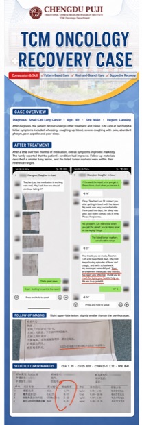
After diagnosis, the patient did not undergo other treatment and chose TCM care at our hospital. Initial symptoms included wheezing, coughing up blood, severe coughing with pain, abundant phlegm, poor appetite and poor sleep.

After treatment

After a little over two months of medication, overall symptoms improved markedly. The family reported that the patient's condition had improved. Follow-up materials described a smaller lung lesion, and the listed tumor markers were within their reference ranges.

Follow-up imaging

Right upper-lobe lesion: slightly smaller than on the previous scan.

Selected tumor markers

CEA 1.70 · CA125 8.07 · CYFRA21-1 2.12 · NSE 8.41. The translated family chat and source evidence panels remain visible in the complete poster.

Case 02 · Xinjiang · Male · Age 71

Lung Cancer After Surgery

A before-and-after report comparison retaining the source measurements, symptom history, examination dates and follow-up findings.

  • Measurements
  • Two report dates
  • Finding comparison
Read the complete English case notes

Before treatment

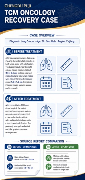
After lung-cancer surgery, follow-up imaging showed multiple nodules in both lungs, some with calcification. The largest nodule near the right oblique fissure measured about 0.6 × 0.4 cm. Multiple enlarged mediastinal and hilar lymph nodes were noted; the largest measured about 1.8 × 1.2 cm. Symptoms included cough, sputum, nausea and dry mouth.

After treatment

After consolidative TCM care at our hospital, the patient reported less cough and sputum. A recent examination described a clear reduction in multiple solid nodules in both lungs, with a trend toward calcification. The previously enlarged mediastinal and hilar lymph nodes were no longer seen.

Source report comparison

30 May 2025: right oblique-fissure nodule about 0.6 × 0.4 cm; mediastinal/hilar lymph node about 1.8 × 1.2 cm. 27 June 2025: multiple solid nodules clearly smaller and trending toward calcification; previously enlarged mediastinal/hilar lymph nodes no longer seen.

Case 03 · Guizhou · Male · Age 59

Lung Cancer With Brain Metastases

A six-month case timeline retaining the presenting symptoms and all three source MRI report dates and findings.

  • 6-month timeline
  • 3 MRI dates
  • Symptom follow-up
Read the complete English case notes

Before treatment

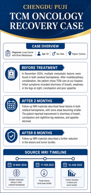
In November 2024, multiple metastatic lesions were found in both cerebral hemispheres. After multidisciplinary consideration, the patient chose TCM care at our hospital. Initial symptoms included shortness of breath, weakness in the legs at night, constipation and poor appetite.

After 2 months

Follow-up MRI materials described fewer lesions in both cerebral hemispheres, with some areas becoming smaller. The patient reported improvement in shortness of breath, constipation and nighttime leg weakness, and appetite returned.

After 6 months

Follow-up MRI materials described a further reduction in the lesions and tumor burden.

Source MRI timeline

15 November 2024: multiple metastatic lesions in both cerebral hemispheres. 11 February 2025: fewer lesions, with some areas smaller. 10 June 2025: further reduction in lesions and tumor burden.

How these cases are prepared

  • The complete source poster and supporting copy are reviewed before English localization.
  • Symptoms reported by the patient or family are kept distinct from findings described in imaging or laboratory materials.
  • Dates, measurements and laboratory values are retained; unclear or unsupported information is not invented.
  • When authentic scan images are not available for the English derivative, clinical icons are used instead of synthetic scans.

Would you like a specialist to review your case?

Send a brief history and your most recent reports. The international care team can help organize the information before a physician consultation.

Small-Cell Lung Cancer · Complete English Poster
Complete English small-cell lung cancer case poster
Lung Cancer After Surgery · Complete English Poster
Complete English lung cancer after surgery case poster
Lung Cancer With Brain Metastases · Complete English Poster
Complete English lung cancer with brain metastases case poster